
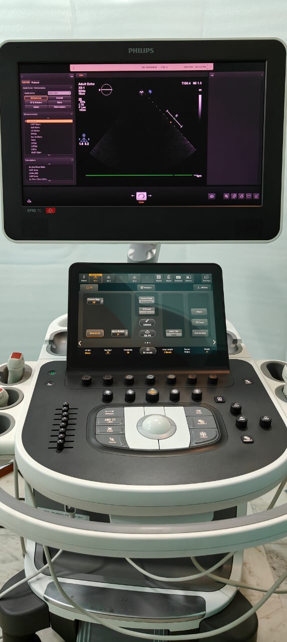
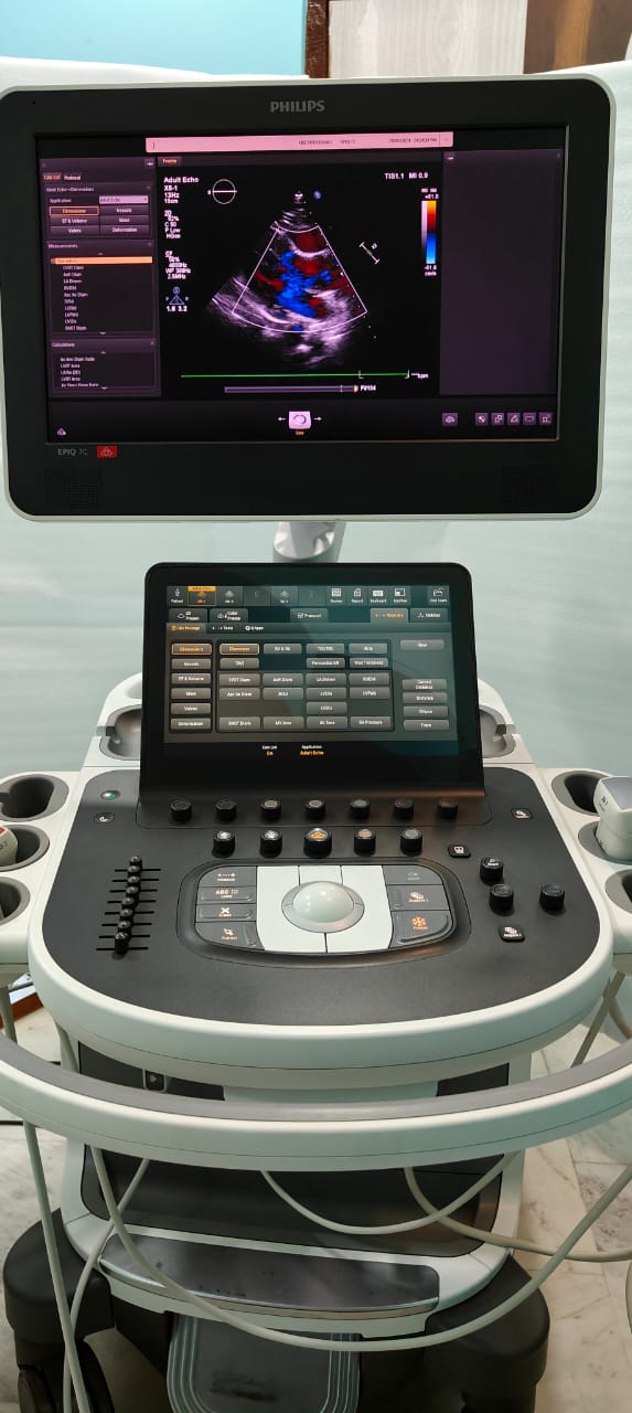
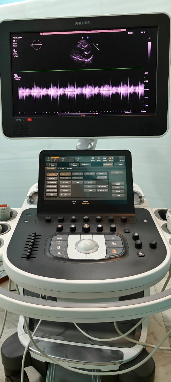
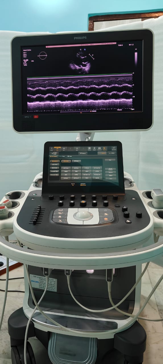
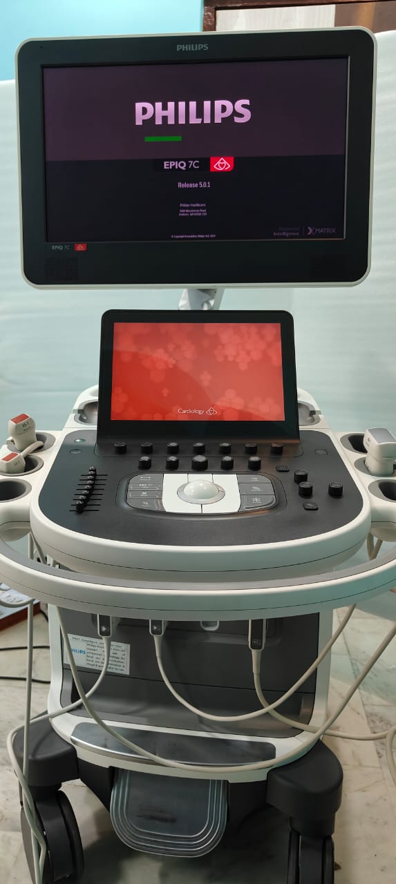
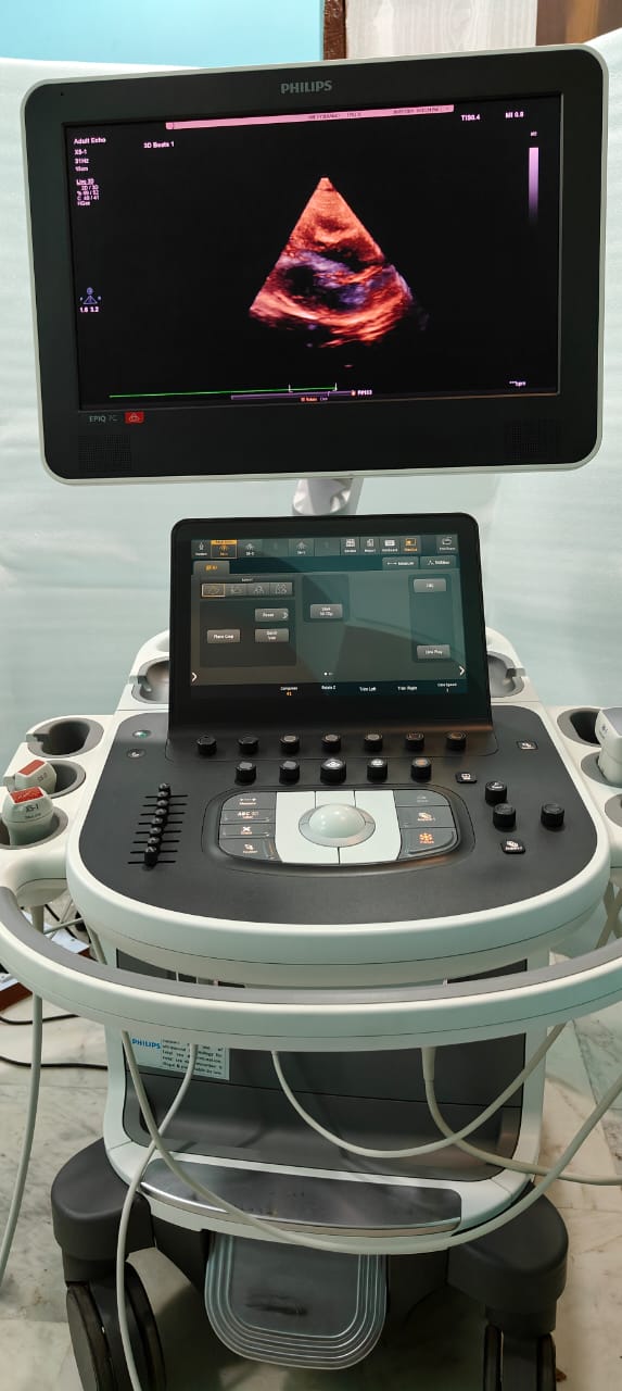
Philips EPIQ 7C Premium Cardiology Ultrasound System, for Cardiac Imaging
WhatsApp us
| Usage/Application |
Cardiac Imaging
|
| Brand |
Philips
|
| Weight |
104.3 kg
|
| Monitor Size |
54.6 cm
|
| Software Technologies |
X Matix
|
| Depth |
109.2 cm
|
| Height |
146 to 171.5 cm
|
| Degree of Movement |
180 degrees
|
| Width |
60.6 cm
|
| Height Adjustment |
25.4 cm
|
Philips EPIQ 7C ultrasound system delivers a new level of clinical confidence with the combination of our most powerful and innovative technologies ever created for ultrasound. Built on key pillars of performance, design, and intelligence. Revolutionary nSIGHT Imaging architecture of EPIQ creates real-time images with precise resolution and exceptional uniformity. EPIQ 7C provides a wide range of enhanced capabilities that redefines premium performance.

Description
Features:
- A wide range of Broadband transducers – catering to all clinical applications.
- PureWave transducers – for enhanced penetration and improved resolution even in difficult to scan patients. Available for both adult and pediatric applications.
- xMatrix Technology – Incorporating one of the most advanced technology with features such as iRotate, iRotate Stress Echo, iCrop, Live X-Plane imaging, 3D & 4D trans-thoracic and trans-oesophageal echo, and even 3D pediatric trans-thoracic echo.
- Mitral Valve Navigator – MVN is designed to take a live 3D volume of the mitral valve and turn it into an easy-to-interpret model in eight guided steps, providing access to a comprehensive list of MV measurements and calculations.
- Anatomical Intelligence (A.I), turning images into answers: (i)a2DQA.I. for AutoEF – Uses A.I. to drive correct region of interest placement on echo view. Speckle tracking assists with maintaining border detection through the cardiac cycle. Once initiated, no user interaction required (ii) aCMQA.I. for GLS and EF – with Zero-click technology – Rapid access to global longitudinal strain from all three apical views.
- Heart Model A.I – This anatomically intelligent cardiac application automatically detects, segments, and quantifies the Left Ventricle (LV) and Left Atrium (LA) from a Live 3D volume.
- HeartModel A.I. provides automated 2D views and reproducible quantification across users and over time, with the workflow efficiency to facilitate faster exams for the precise measurement of cardiac function.
- 3D TTE and TEE – Fast, high-quality 3D trans-thoracic and trans-esophageal imaging, with quick access to full volume 3D from 2D images, providing a high level of diagnostic confidence.
- Environmentally responsible ultrasound – 25% less power consumption than previous generations of premium platforms.
- “Library-quiet” operation – EPIQ is almost silent when running, with a decibel range of 37-41db, which is equivalent to the sound levels in a library!
Reviews (0)
Reviews
There are no reviews yet.


-
(0 customer reviews)